Corneal epithelium

Results: 15


#Item
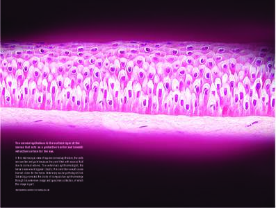
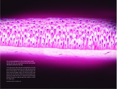
1The corneal epithelium is the surface layer of the cornea that acts as a protective barrier and smooth refractive surface for the eye.  In this microscopic view of equine corneal epithelium, the cells are swollen and pa

The corneal epithelium is the surface layer of the cornea that acts as a protective barrier and smooth refractive surface for the eye.  In this microscopic view of equine corneal epithelium, the cells are swollen and pa

Add to Reading List

Source URL: vision.wisc.edu

Language: English - Date: 2016-03-04 11:57:34
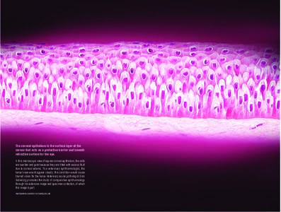
    2The corneal epithelium is the surface layer of the cornea that acts as a protective barrier and smooth refractive surface for the eye.  In this microscopic view of equine corneal epithelium, the cells are swollen and pa

    The corneal epithelium is the surface layer of the cornea that acts as a protective barrier and smooth refractive surface for the eye.  In this microscopic view of equine corneal epithelium, the cells are swollen and pa

    Add to Reading List

    Source URL: www.vision.wisc.edu

    Language: English - Date: 2016-03-04 11:57:34
      3The corneal epithelium is the surface layer of the cornea that acts as a protective barrier and smooth refractive surface for the eye.  In this microscopic view of equine corneal epithelium, the cells are swollen and pa

      The corneal epithelium is the surface layer of the cornea that acts as a protective barrier and smooth refractive surface for the eye.  In this microscopic view of equine corneal epithelium, the cells are swollen and pa

      Add to Reading List

      Source URL: vision.wisc.edu

      Language: English - Date: 2016-03-04 11:57:34
        4ETHZ-JST Joint WS  Abstract of Presentation Presentation Title: Corneal reconstruction using tissue-engineered epithelial cell sheets fabricated ex vivo from autologous oral mucosal epithelium

        ETHZ-JST Joint WS Abstract of Presentation Presentation Title: Corneal reconstruction using tissue-engineered epithelial cell sheets fabricated ex vivo from autologous oral mucosal epithelium

        Add to Reading List

        Source URL: www.jst.go.jp

        - Date: 2013-09-02 04:06:04
          5

          PDF Document

          Add to Reading List

          Source URL: eyefreedom.com

          Language: English - Date: 2009-06-23 12:42:01
          6Cross-linking procedure UV-X™ 1000 Indications  Progressive keratoconus  Iatrogenic ectasia (e.g. after LASIK)  Pellucid marginal degeneration

          Cross-linking procedure UV-X™ 1000 Indications  Progressive keratoconus  Iatrogenic ectasia (e.g. after LASIK)  Pellucid marginal degeneration

          Add to Reading List

          Source URL: www.medsrl.com.ar

          Language: English - Date: 2014-05-14 11:39:45
          7Cross-linking procedure UV-X™ 2000 Indications  Progressive keratoconus  Iatrogenic ectasia (e.g. after LASIK)  Pellucid marginal degeneration

          Cross-linking procedure UV-X™ 2000 Indications  Progressive keratoconus  Iatrogenic ectasia (e.g. after LASIK)  Pellucid marginal degeneration

          Add to Reading List

          Source URL: www.medsrl.com.ar

          Language: English - Date: 2014-05-14 11:39:51
          8Microsoft Word - ESCRS2010ComplicationsCourseHandout.doc

          Microsoft Word - ESCRS2010ComplicationsCourseHandout.doc

          Add to Reading List

          Source URL: lasikscandal.com

          Language: English - Date: 2013-04-13 04:29:23
          9To our knowledge, orbital myositis has only once been previously reported.1 In conclusion, we present a case of superior rectus muscle myositis due to CSS. The concurrent association with an eosinophilic conjunctival nod

          To our knowledge, orbital myositis has only once been previously reported.1 In conclusion, we present a case of superior rectus muscle myositis due to CSS. The concurrent association with an eosinophilic conjunctival nod

          Add to Reading List

          Source URL: archopht.jamanetwork.com

          Language: English
          10Table 3. CNV Lesion Size and Last FA Results Total Area of CNV, mm2 Patient

          Table 3. CNV Lesion Size and Last FA Results Total Area of CNV, mm2 Patient

          Add to Reading List

          Source URL: archopht.jamanetwork.com

          Language: English